Sometimes, but a CT scan is best at showing bone. Many of the problems that cause daily back pain involve soft tissue, such as a disc or a nerve, and an MRI usually shows those more clearly.
I am Dr. Tony Mork, MD, a board-certified orthopedic surgeon. I have practiced endoscopic spine surgery exclusively since 1998, and I have performed more than 8,000 endoscopic spine procedures. Here is how I explain scans to patients who sit in my office after months of pain, wondering why no one has found the cause.
TL;DR
- What a CT does well: It shows bone in fine detail, including fractures, bone spurs and the shape of the spine before or after surgery.
- What it can miss: It is less sensitive than an MRI (magnetic resonance imaging) for discs, nerves and ligaments, which are common sources of pain.
- When it is used: Most new back pain does not need a scan right away. A CT is often chosen when an MRI cannot be done, or when the MRI leaves questions.
- If your scan is normal: A normal scan does not mean your pain is imagined. An exam and an MRI are needed to know for sure, and accurate diagnosis comes before any surgery.
- Surgery is not the first step: Many back problems improve with time and conservative care. Fusion is a last resort that can still be done later if needed.
When does a doctor order a CT scan for back pain?
A doctor usually orders a CT (computed tomography, a series of x-ray images assembled into cross-sections of your body) when a question about bone needs an answer, or when an MRI is not possible.
Most back pain does not start that way. Sudden pain after lifting a bag of mulch or sitting through a long drive often settles with time, activity changes and gentle movement. According to the American College of Radiology’s appropriateness criteria, a CT of the lower spine is rated “usually not appropriate” as the first image for new low back pain with no warning signs. Guidelines from the North American Spine Society also say there is not enough evidence to recommend for or against imaging in that situation.
So a scan is not a test you need on day one. It becomes useful when:
- Pain lingers: You have had symptoms for many weeks despite sensible care.
- Nerve symptoms appear: You have leg burning, numbness or weakness, or sciatica (pain that runs from the low back down the leg).
- Injury occurred: A fall, a car accident or a heavy lifting injury raises the question of a fracture.
- You cannot have an MRI: A pacemaker or an older implant may rule it out.
- Surgery is being planned: Surgeons often want to see bone detail before an operation.
If you have already had spine surgery, a CT can also show how the bone and any hardware are sitting.
What can a CT scan show in your spine?
A CT scan shows bone in exceptional detail. That makes it very good at finding fractures, bone spurs, joint wear, tumors that affect bone and structural differences you were born with.
Johns Hopkins Medicine notes that a spine CT may be used to look for a herniated disc, tumors, the extent of an injury and other structural problems, particularly when an x-ray or exam does not settle the question. The images are built in slices and then reformatted, so your doctor can look at the spine from the side and from the front.
That detail helps in specific cases:
- Facet joints: These are the small joints at the back of the spine. A CT shows arthritis and overgrowth of bone there, which can be part of facet syndrome (pain from irritated or worn facet joints).
- Bone spurs: A CT shows whether bone has grown into the space where a nerve travels.
- Fractures: A CT shows small breaks that an x-ray can miss.
- Calcified tissue: A 2020 review in the journal literature on spine imaging reports that CT is better than MRI at separating hardened tissue from surrounding soft tissue.
The opening for a nerve is called the foramen. When bone narrows it, that is foraminal stenosis (narrowing of the opening where a nerve leaves the spine). A CT can show the bony part of that narrowing well. You can read how I treat it on my page about foraminal stenosis treatment.
What can a CT scan miss?
A CT scan can miss problems in soft tissue, including many disc problems, and it cannot show most nerve irritation directly.
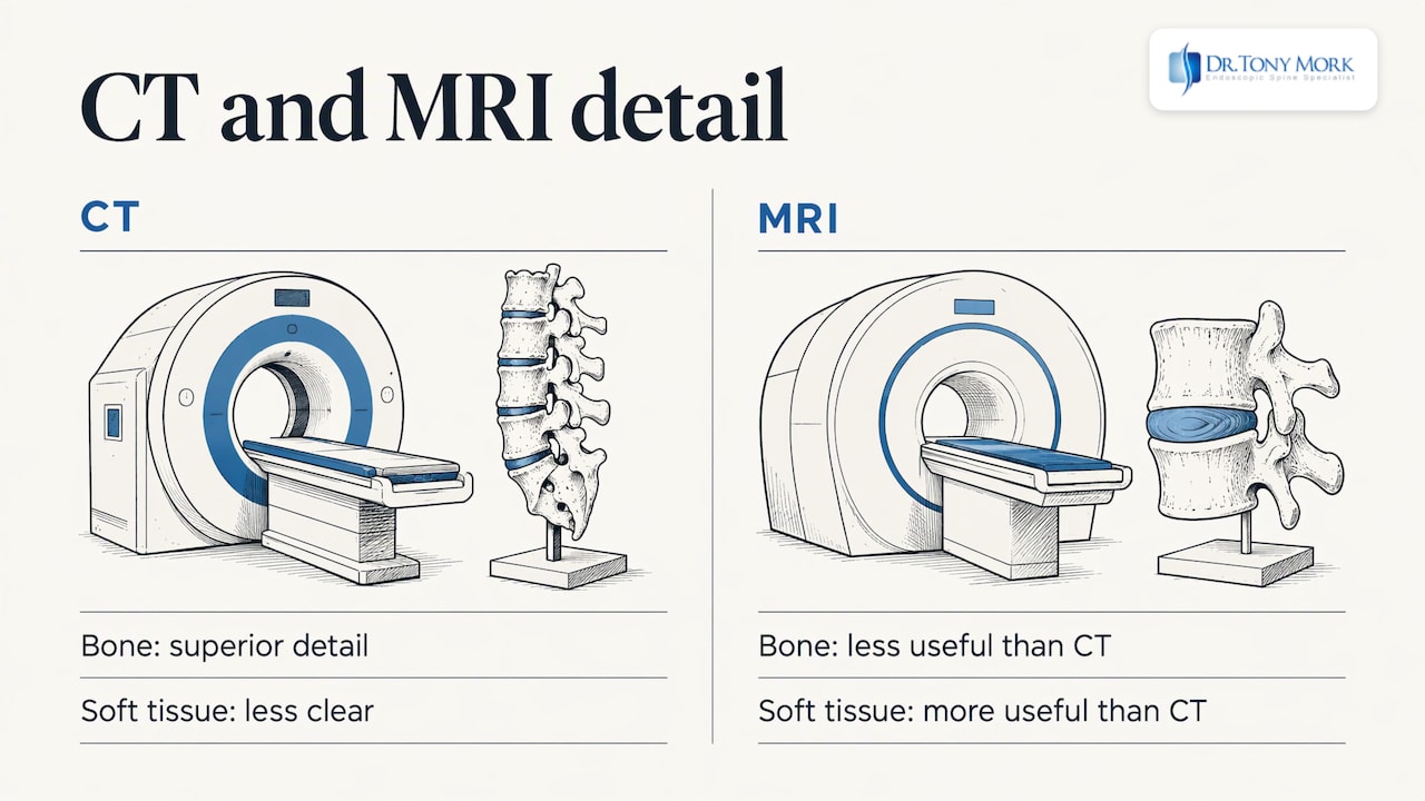
Here is the picture I use with patients. Think of a disc as a liquid-filled cushion between two vertebrae (the stacked bones of your spine). It has a tough outer wall and a softer center. When the wall tears or bulges, the cushion no longer works the way it was designed to. A CT shows the bones on either side of the cushion very well. It shows the cushion itself, and the small tears in its wall, far less clearly.
The World Federation of Neurosurgical Societies’ spine committee (2024) makes the same point: CT is superior for bone, but not as useful as MRI for soft-tissue problems such as disc disease or spinal stenosis (narrowing of the space around the nerves). The 2020 review I mentioned says CT is less sensitive for the nerves and ligaments.
Position matters too. You lie flat for a CT. Pain that comes on when you sit, stand, drive or lift a grandchild may not show up well when you are lying down, because the spine carries load differently.
That is one reason people with a deep ache in the low back that gets worse in the car can leave a CT with a report that says very little. The scan was accurate for what it can see. It was not designed to see the source of your pain.
CT scan or MRI: which is better for back pain?
For most back pain, MRI is the better first choice, because it shows discs, nerves and soft tissue without ionizing radiation (the type of radiation used in x-rays and CT). A CT is the better tool for bone.
A 2021 appropriateness update from a provider-led group says most guidelines prefer MRI for low back pain for those reasons. It lists CT as appropriate when MRI cannot be done, when MRI findings are unclear or do not match your symptoms, for surgical planning or fusion evaluation, and for bone abnormalities such as fractures.
| Question | CT scan | MRI |
|---|---|---|
| Best at showing | Bone, fractures, bone spurs | Discs, nerves, ligaments, soft tissue |
| Radiation | Yes, a small amount per scan | None |
| Time | A few minutes | Usually longer, in a tube that is louder and tighter |
| Metal implants | Usually fine | May rule it out or blur the image |
| Annular tears and disc bulges | Limited | Often visible |
| Planning surgery | Useful for bone detail | Useful for nerve and disc detail |
If you have a pacemaker or an older implant that is not MRI compatible, a CT-based test may be the right path. Your doctor can tell you which implants allow an MRI.
If you only have a CT report and still have pain, I would want to see an MRI as well. In my practice I review MRI and CT images remotely, which helps patients who live far away or out of state.
What if my scan is normal but I still hurt?
A normal scan does not mean nothing is wrong. It means the test did not find a structural reason for your pain, and that is a reason to look further, not to give up.
I hear this often. A person has seen three or more doctors. Their hips were checked. A hernia exam was normal. The MRI was called normal. Yet they still have deep midline low back pain, groin or tailbone pain, or a back that “goes out” again and again. Pain does not have to match the picture on a standard scan. A disc can hurt because of a small tear in its outer wall or a tiny area of inflammation that a quick read can pass over.
Things I look for in a careful review:
- Annular tear: A crack in the disc wall (see annular tear treatment for how I approach disc problems).
- Disc bulge: A disc that pushes outward without fully breaking through.
- High intensity zone (HIZ): A bright spot on an MRI in the disc wall that can signal a painful tear.
- Pain on the opposite side: A herniated disc can sit on one side while the pain shows up on the other.
- Facet joints: Joint pain that mimics a disc problem.
I also look at how your pain behaves. Pain that worsens when you sit, eases when you stand, and returns after physical therapy or short-lived injections tells me something about its source.
Some patients ask about a CT discogram. That test places dye in a disc and then uses a CT to look at the disc and see whether the pressure reproduces your pain. It is one way to link a disc to your symptoms. It is not for everyone, and it is only one piece of a diagnosis alongside your history, your exam and your MRI.
I cannot diagnose you from an article, and nobody should. An exam and an MRI are needed to know for sure.
What happens during a back CT scan?
You lie on a table that slides through a ring-shaped scanner, and the scan itself takes only a few minutes. According to the Florida Agency for Health Care Administration’s HealthFinder, a lumbosacral spine CT usually lasts a few minutes, and you need to stay still because movement blurs the pictures.
Here is the usual routine:
- Before: You remove metal such as a belt or jewelry and may change into a gown.
- Contrast: In some cases, a dye containing iodine is given through a vein to make certain tissues stand out. Tell the staff about any allergy to dye or any kidney problem.
- Scan: You lie still, sometimes on your back, and may be asked to hold your breath briefly.
- After: Without contrast, most people go back to normal activity and diet unless the doctor says otherwise. Johns Hopkins Medicine notes that if contrast is used, you may be watched for a period for itching, swelling, rash or trouble breathing.
If lying flat is painful, say so before you start. A pillow under your knees often helps, and the staff can work with you.
Is the radiation from a CT scan a concern?
The radiation from one CT is small, but it adds up when scans are repeated, so a scan should answer a real question.
The Florida HealthFinder says scans are tightly monitored to use the least radiation needed, that the risk from any single scan is small, and that the risk rises as the number of scans grows. The American College of Radiology lists a relative radiation level of 1 to 10 mSv (millisieverts, a unit of radiation dose) for a lumbar spine CT without contrast. A practice parameter from several radiology societies also says protocols should be tailored to the reason for the scan, to balance image quality and dose.
In practice:
- Keep your records: Bring prior scans and reports so no one repeats a test you already had.
- Ask about the question: Know what your doctor wants the scan to answer.
- Pregnancy: If you might be pregnant, tell the staff before any scan.
I would rather review the images you already have than add another scan that does not change the plan.
Which back pain symptoms need urgent care instead of a scan?
Some symptoms mean you should seek emergency care now, not wait for imaging. Go to an emergency room if you have back pain with:
- Loss of bladder or bowel control: Or trouble urinating.
- Numbness in the saddle area: The groin, inner thighs and buttocks.
- Progressive leg weakness: Weakness that worsens over hours or days.
- Fever with severe back pain: Particularly if you recently had an infection or procedure.
- Back pain with a history of cancer, or after a significant injury: Seek prompt evaluation.
The Cleveland Clinic Journal of Medicine describes cauda equina compression (pressure on the bundle of nerves at the base of the spine) as classically involving back pain, sciatica on both sides, saddle numbness and leg weakness. When it is suspected, an MRI is the study of choice and the evaluation should be immediate.
These problems are rare. Serious causes affect fewer than 1% of people with low back pain, according to a 2025 imaging guideline for lumbar MRI. Still, they are the situation where speed matters most. Seeing an emergency physician comes first.
How do I know if I need surgery or a fusion?
Most people do not need a fusion, and many do not need surgery at all. Time is on your side in many cases.
I always start with conservative care when it fits: time, activity changes, physical therapy, medication and injections such as an ESI (epidural steroid injection, a shot of medicine near the irritated nerve). Many back problems improve with these steps. If you are on medication, keep taking it as your doctor directs, and do not stop it without asking them.
Surgery enters the conversation when pain continues, the cause is clear, and daily life is shrinking. Sitting through a workday, putting on shoes, sleeping or playing golf becomes a struggle. Then the goal is to remove the source of pain, not only to manage it.
The spine was designed to move. A fusion joins vertebrae so they no longer move. I consider fusion a last resort, and it can still be done later if it is ever needed. That is why I try to avoid taking that option off the table early. If you were told fusion is your only choice, I would want to look at your images before you decide. See my page on minimally invasive surgery for the larger picture.
Here is how I describe endoscopic spine surgery: a small incision, a camera, outpatient, no hardware, no fusion. “Minimally invasive” refers to how much healthy tissue is disturbed, not only the size of the cut. A laser may be used as one tool during the operation. It is not a cure by itself.
Honest pros and cons matter, so here they are:
- Benefits: Less disturbance of healthy tissue, outpatient care, and no hardware. Many patients return to regular activities sooner than after open surgery. Results vary.
- Recovery: Recovery often takes some days to some weeks depending on the person and the problem.
- Limits: Not every spine problem is a fit for an endoscope. Severe deformity or instability may need a different approach. Any surgery carries risks such as infection, bleeding or symptoms that persist.
- Diagnosis first: The operation only helps when it targets the right source, which is why accurate diagnosis comes before surgery.
You can read more on my pages for endoscopic spine surgery, herniated disc treatment and spinal stenosis treatment.
What do I tell patients about scans?
I founded the Endoscopic Spine Academy and co-founded Microspine, and I have spent my career on one question: where does this person’s pain come from?
A scan is a picture. It is not a diagnosis. I have seen patients with dramatic images and little pain, and patients with quiet images and a great deal of pain. That is why I read images alongside your story and your exam.
If you have a CT, I want the MRI too. If you have had an epidural injection that helped for a short time, or therapy that made things worse, or a chiropractor whose relief never lasts, those details matter. They are clues about the source.
My practice is built so that you can reach me directly, before and after surgery. I want you to understand your options, including the ones that do not involve an operation.
Dr. Tony Mork
Can you send me your scans for review?
Yes. I offer remote evaluation of MRI and CT images, and I also offer phone appointments, so you can talk through your options without travel. My patients come from Orange County and Southwest Florida, and many travel from out of state.
You can find me at my Irvine office, 2102 Business Center Dr #127, Irvine, CA 92612, and at my Naples, Florida office. Insurance may cover facility and anesthesia fees, while surgeon’s fees are paid directly. Start at my concierge spine services page.

FAQ
Is a CT scan or an MRI better for back pain?
For most back pain, an MRI is better because it shows discs, nerves and soft tissue clearly and uses no radiation. A CT is better for bone. Your doctor may choose a CT if you cannot have an MRI or if the MRI leaves a bone question open.
How long does a CT of the lower back take?
The scan itself usually takes a few minutes, according to the Florida HealthFinder. Plan for more time overall for check-in, changing and any contrast dye. Results usually come from a radiologist’s written report afterward.
Can a CT scan show a herniated disc?
Sometimes. A CT can suggest a herniated disc, and Johns Hopkins Medicine lists it as one reason for the test. An MRI shows the disc and the nerve more clearly, so I rely on it when I judge a disc problem.
What should I do if my CT report says everything is normal?
Keep your images and report, and ask for an MRI if you have not had one. A normal CT does not rule out a disc tear or a nerve problem. If you want a second opinion, I can review your images remotely.
If you have a CT or an MRI, upload it through my office and I will review it personally and talk with you about your options.
This article is for general education and is not medical advice. Please see a qualified doctor about your symptoms.