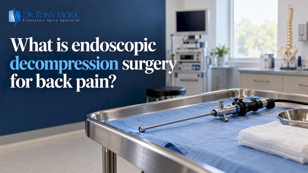
Endoscopic decompression is outpatient surgery that frees a pinched spinal nerve through a small incision, using a camera and thin instruments. It removes the disc, bone, or ligament pressing on the nerve, and it does not require a fusion or hardware.
I am Dr. Tony Mork, a board-certified orthopedic surgeon. I have practiced endoscopic spine surgery exclusively since 1998 and have performed more than 8,000 endoscopic spine procedures. If you are worried you will need a fusion, this page explains your options in plain English.
TL;DR
- What it is: a small incision, a camera, and fine instruments that remove tissue pressing on a nerve.
- Who it may help: people whose herniated disc or stenosis (narrowing around the nerves) is confirmed by an exam and an MRI (magnetic resonance imaging).
- What it avoids: a large muscle-cutting incision, hardware, and fusion in most cases.
- What comes first: an accurate diagnosis, and conservative care where it fits. Many back problems improve with time.
- What to expect: many patients go home the same day. Recovery often takes weeks, and results vary.
What is endoscopic decompression, and how does it work?
Endoscopic decompression means taking pressure off a nerve while looking at it through a camera. “Decompression” is the goal. “Endoscopic” describes the tool: an endoscope, a thin tube with a camera and light on the end.
Think of a garden hose with a kink or a heavy object sitting on it. The water cannot flow. A nerve squeezed by a disc fragment or overgrown bone behaves the same way. You may feel it as burning down the leg, numbness in the foot, or pain that makes it hard to put on your shoes.
Through an incision of roughly a centimeter, I pass the endoscope and small instruments to the problem area. The American Academy of Orthopaedic Surgeons (AAOS) describes endoscopic discectomy as using a camera-equipped endoscope through small incisions to remove the disc portion that compresses the nerves. I can see the nerve directly on a screen and remove only what is pressing on it.
In my practice, the procedure comes down to five features:
- Incision: small, often about the size of a fingertip.
- Camera: a magnified view of the nerve and the tissue pressing on it.
- Setting: outpatient, so you usually go home the same day.
- Hardware: none. No screws or rods are placed.
- Fusion: none. The spine keeps its motion.
Why does “minimally invasive” matter if the incision is the only difference?
It does not refer only to incision size. Minimally invasive describes how much healthy tissue is disturbed to reach the problem. A short scar can still hide a lot of muscle stripped off the bone underneath.
Endoscopic work passes through a narrow channel and gently separates tissue rather than cutting through it. The muscles that hold your spine upright stay largely intact. That matters when you want to get back to sitting through a workday or lifting a grandchild. You can read more in my article on how minimal minimally invasive spine surgery can get.
Which conditions can endoscopic decompression treat?
It is used mainly for two groups of problems: herniated discs and spinal stenosis. A peer-reviewed 2019 review of endoscopic decompression techniques identifies these as the two principal disease groups. An exam and an MRI are needed to know for sure what is behind your symptoms.
The conditions I treat this way include:
- Herniated disc: the soft center of a disc pushes out and presses on a nerve. The disc works like a liquid-filled cushion, and a herniation is a leak through the wall. See my page on endoscopic discectomy for a herniated disc.
- Spinal stenosis: narrowing of the central canal that holds the nerves. People often notice leg heaviness or pain after standing or walking, eased by sitting or leaning on a shopping cart. See endoscopic decompression for spinal stenosis.
- Foraminal stenosis: narrowing of the opening where a nerve leaves the spine. It can cause sciatica in the lumbar spine or arm symptoms in the neck. See endoscopic foraminal decompression.
- Facet syndrome: painful, arthritic facet joints (the small joints at the back of the spine). Diagnosis is more subtle, as I explain below.
- Bulging discs and annular tears: some cause enough nerve irritation to need treatment. Many do not. See my page on bulging disc treatment.
- Pain after earlier spine surgery: sometimes a nerve is still compressed or a source of pain was missed.
When I say sciatica (pain traveling from the low back down the leg), I mean a symptom, not a diagnosis. AAOS explains that sciatica comes from compression of a lumbar nerve root, commonly by a herniated disc or spinal stenosis. Finding which one applies to you is the real work.
Should I try conservative care before surgery?
Yes, where it fits. Many back problems improve with time, activity changes, physical therapy, medication, and sometimes injections. Time is on your side in most cases.
AAOS notes that surgery for sciatica may be considered when disabling leg pain persists after 3 or more months of nonsurgical treatment. For lumbar stenosis, AAOS says surgery is generally reserved for people whose pain or weakness substantially reduces quality of life, such as difficulty walking for extended periods.
Some situations should not wait. Tell a doctor right away if you notice new leg weakness, loss of bladder or bowel control, or numbness in the groin or saddle area. Those can signal a serious nerve problem needing urgent care.
I would never ask you to stop a medication or skip your own doctor. If you have tried a reasonable course of care and the pain keeps returning, that is the time to look for the source.
Why does an accurate diagnosis have to come first?
Because surgery only works when it targets the actual source of pain. My goal is to remove that source, not just manage the symptoms. If the wrong structure is treated, you can have an operation and still hurt.
A good diagnosis combines your story, a physical exam, and imaging such as an MRI or a CT (computed tomography) scan. AAOS describes the exam and imaging tests as how lumbar spinal stenosis is assessed and confirmed.
Some sources of pain are harder to pin down. Facet pain is the clearest example. A published consensus guideline on lumbar facet interventions reports that history, exam, and imaging alone do not reliably identify it, and that a diagnostic block (a numbing injection to test a suspected pain source) is more dependable. I explain this further in my articles on diagnosing facet syndrome and diagnosing foraminal stenosis.
If you have been told your MRI is normal but you still hurt, or you have seen several doctors without an answer, the image still deserves a careful second look matched against your symptoms.
What happens during endoscopic decompression surgery?
You are positioned on your stomach or side, the area is numbed, and I work through a small channel to reach the nerve. Anesthesia is tailored to you and your procedure.
The path to the spine depends on where the problem sits:
- Transforaminal route: from the side, through the opening where the nerve exits. It is often used for disc herniations and foraminal stenosis. See my article on the lumbar transforaminal approach.
- Interlaminar route: from the back, between two vertebrae (the bones of the spine). It is often used for central stenosis.
A published 2025 review of endoscopic lumbar surgery describes removal of disc material, bone, or thickened ligament and facet tissue under direct view, with constant irrigation to keep the field clear. I use a laser in some cases. It is one tool among several, never a cure by itself. The cure comes from correctly identifying and removing what presses on the nerve.
The incision is closed with a stitch or a small bandage. After a short recovery period, most patients go home the same day.
How is it different from open decompression and spinal fusion?
Endoscopic decompression disturbs less healthy tissue than open surgery, and unlike fusion it does not join any bones. The spine was designed to move. I try to protect that motion whenever the problem allows.
| Endoscopic decompression | Open decompression | Spinal fusion | |
|---|---|---|---|
| Goal | Remove what presses on the nerve | Remove what presses on the nerve | Stabilize and join vertebrae |
| Incision | Small | Larger | Larger |
| Muscle disruption | Limited | More | More |
| Hardware | None | Usually none | Screws, rods, or cages |
| Motion | Preserved | Preserved | Lost at the fused level |
Fusion is a last resort. It can still be done later if needed, so choosing endoscopic surgery first does not close that door. AAOS notes fusion may be recommended when arthritis has progressed to spinal instability, meaning a segment that slips or moves abnormally. That is a real indication, and decompression does not stabilize anything on its own.
When a surgeon recommends fusion for a pinched nerve without instability, I believe it deserves a second opinion. You can read more on my page about the pros and cons of endoscopic spine surgery.
What are the honest pros and cons?
Endoscopic decompression offers meaningful advantages, along with real limits. You should hear both.
Advantages
- Less tissue disturbed: muscles and bone are largely preserved.
- Outpatient care: many patients go home the same day.
- Motion preserved: no hardware and no fusion.
- Research: a 2021 meta-analysis in the Journal of Neurosurgery: Spine reported shorter hospital stays and lower postoperative back pain scores with endoscopic methods than with other minimally invasive approaches for lumbar stenosis.
Limits
- Not for every spine: severe instability, a deformity, or a need for extensive exposure may call for a different operation, including fusion.
- Selection matters: a 2021 review lists severe fibrotic scarring, a heavily calcified disc, and cauda equina syndrome (compression of the nerve bundle at the base of the spine, an emergency) as situations outside the usual indications.
- Skill-dependent: the technique has a learning curve, so surgeon experience matters. I founded the Endoscopic Spine Academy and co-founded Microspine to teach and refine these methods.
- No guarantees: incomplete relief, recurrent herniation, and nerve irritation can occur. Results vary.
How long does recovery take?
Recovery often takes a few weeks, though it varies by condition, age, and job. I cannot give you an exact timeline, and I will not promise immediate relief or a pain-free result.
Many patients report walking the same day and returning to light daily activities within days. Heavier lifting, golf, and training usually come later. Nerves can take weeks to settle after pressure is removed, so numbness or tingling may linger before it fades.

In my concierge practice you have direct access to me after surgery, so questions get answered quickly. You can find more in my endoscopic spine surgery guide for patients.
What does my practice offer for endoscopic decompression?
I see patients at my Irvine office, 2102 Business Center Dr #127, Irvine, CA 92612, and at my Naples, Florida office. Many patients also travel from other states. If you cannot visit first, I can review your MRI or CT scan remotely.
You can learn how endoscopic spine surgery works in my practice, or start with an imaging review through my concierge spine services. Insurance may cover facility and anesthesia fees, but not the surgeon’s fee, and payment is arranged before surgery.
What is my view on decompression versus fusion?
Most patients who come to me have been told, directly or by implication, that fusion is where their story ends. I disagree with routine fusion. Fusion removes motion, and the spine was designed to move.
My approach is to find the true source of pain first, through the exam, the images, and sometimes diagnostic injections. Then I remove that source through the smallest path that lets me see it, with no hardware and no fusion. The goal is to remove what causes the pain, not to manage it indefinitely.
Endoscopic surgery has limits, and I will tell you when it is not right for you. Fusion remains available later if it is ever needed. Choosing the gentler option first costs you nothing in that respect.
Dr. Tony Mork
Frequently asked questions
Is endoscopic decompression the same as a laminectomy?
A laminectomy removes part of the lamina, the bony roof over the spinal canal, to give nerves room. Endoscopic decompression can include this step, done through a small channel with a camera instead of a large open incision. The aim is the same: take pressure off the nerve.
Can endoscopic surgery treat spinal stenosis?
It can for many people with imaging-confirmed stenosis causing leg or back symptoms. Candidates are usually those whose symptoms have not improved with conservative care. Severe instability or deformity may need another approach. An exam and an MRI are needed to know for sure.
Can endoscopic surgery help facet syndrome?
Sometimes. Facet pain is hard to confirm from imaging alone, so I rely on the exam and often a diagnostic block before recommending treatment. Surgery makes sense only when the facet joint is confirmed as the source.
Will I need a fusion later?
Not necessarily. Many patients never need one. If a problem such as instability develops, fusion can still be done later, so having endoscopic surgery first does not rule it out.
If you have an MRI, upload it and I will review it personally and talk with you about your options. You can request a consultation here.
Recommended
- Endoscopic Spine Surgery Guide For Patients
- Endoscopic Spine Surgery
- The Pros and Cons of Endoscopic Spine Surgery
- How is discogenic back pain treated?
This article is for general education and is not medical advice. Please see a qualified doctor about your symptoms.